

Akshay Arun Vetal1 , Rachita S. Dhurat1 , M. V. Chethan1 , Darshan Jain1 1 Department of Dermatology, Lokmanya Tilak Municipal Medical College and General Hospital, Mumbai, Maharashtra, India
ABSTRACT
Incomplete split earlobes, frequently resulting from heavy and pendulous ear jewelry, can cause significant
cosmetic and psychological distress. Traditional lobuloplasty techniques often face challenges, particularly in
achieving precise and symmetrical results. is study introduces an innovative lobuloplasty technique using a
customized elliptical biopsy punch to repair Type I and Type IIA earlobe deformities. By modifying a standard circular
biopsy punch, we achieved better margin symmetry and tissue approximation. e procedure involves a two-stage
process of stamping and circumduction to excise the defect, followed by closure with vertical mattress sutures. is
technique offers several intraoperative advantages, including reduced tissue handling, minimized blood loss, and time
efficiency, leading to superior cosmetic outcomes. Our findings suggest that this method is a significant improvement
over conventional techniques, providing a more user-friendly approach and enhanced patient satisfaction.
Keywords: Elliptical biopsy punch, Types of earlobe tear, Lobuloplasty, Time saving, Better cosmetic outcomes
*Corresponding author: Akshay Arun Vetal, Department of Dermatology, Lokmanya Tilak Municipal Medical College and General Hospital, Mumbai, Maharashtra, India
Received: 24 July 2024
Accepted: 15 August 2024
Published: 24 September 2024
Published:
DOI
10.25259/JCAS_49_2024
Videos available on:
https://doi.org/10.25259/
JCAS_49_2024
PROBLEM STATEMENT
Earlobe piercing is a common practice worldwide. In the Indian subcontinent, it has deep cultural
roots. It is also used as a fashion statement and a form of self-expression in modern times.
A common complication of earlobe piercing is the eventual widening of the ear hole. Common
causes include the usage of heavy and pendulous ear jewelry. Chronic traction on the earlobe by
the jewelry results in atrophy of tissue and gradual widening of the ear hole, subsequently leading
to a split earlobe.1 is problem often compels patients to avoid wearing small ear studs as they
may not fit well and fall off.
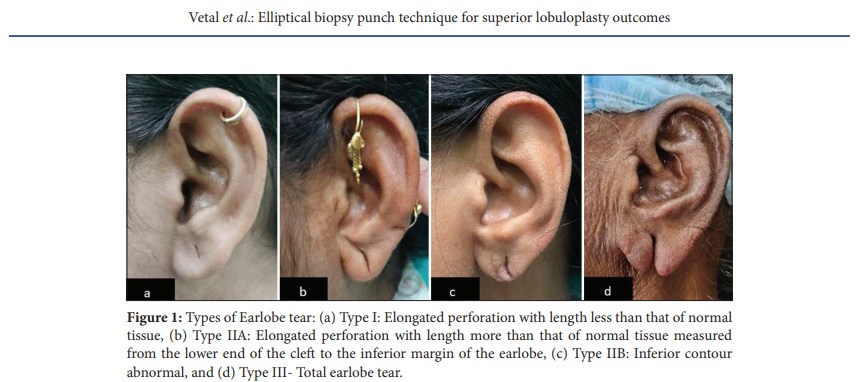
Sadasivan and Kochunarayan proposed a revised classification of split earlobe deformity in 20202
[Figure 1]:
- Type I: Elongated perforation with a length less than that of normal tissue
- Type IIA: Elongated perforation with length more than that of normal tissue measured from
the lower end of the cleft to the inferior margin of the earlobe - Type IIB: Inferior contour abnormal, ptosis of earlobe present
- Type III: Total earlobe tear.
The earlobe is one of the most fleshy and mobile areas on the body, making it difficult to keep
steady during cosmetic repair. Maintaining linear cuts is challenging, especially with the earlobe’s
tendency to slip during handling. For Type I and Type IIA earlobe tear repair, traditional methods
This is an open-access article distributed under the terms of the Creative Commons Attribution-Non Commercial-Share Alike 4.0 License, which allows others
to remix, transform, and build upon the work non-commercially, as long as the author is credited and the new creations are licensed under the identical terms.
©2024 Published by Scientific Scholar on behalf of Journal of Cutaneous and Aesthetic Surgery
often involve freshening the cleft margins by excising cleft
tissue with a scalpel.3
However, incomplete excision or overexcision can result in grooving along the suture line, notching
of the inferior margin, and contour irregularities. Creating
equal margins on both sides of the cleft is challenging. In
addition, this method is time-consuming, as incisions are
required over the lateral as well as the medial aspect of the
earlobe. Maintaining hemostasis is another challenge due to
the time-consuming nature of the procedure.
RECOMMENDED SOLUTION
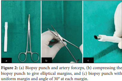
In this modified technique, we converted a traditional
circular 5 mm biopsy punch into an elliptical one [Figure 2].
e biopsy punch was compressed on both ends using
artery forceps to create an elliptical cutting edge with
two symmetrical arcs that intersected at an angle of 30° or
less [Video 1]. Depending on the length of the Type I and
Type IIA earlobe, a punch of a smaller or larger size was
used. e earlobe was held in place using a chalazion clamp.
e flat surface was placed against the medial aspect of
the earlobe, and the ring was placed over the lateral aspect
such that the ear hole lay in the center. e clamp was then
tightened.
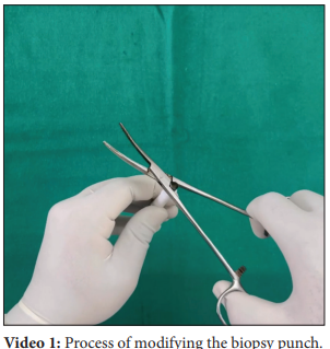
The innovative technique involved two main stages: Stamping
and circumduction movement [Video 2]. e initial step
involved positioning the oval punch over the enlarged ear
hole and applying firm, even pressure, to create an imprint.
is “stamp” marked the boundaries of the area to be excised,
providing a guide for the next step. Following the initial
imprint, the punch was carefully rotated by a circumduction
movement while maintaining pressure. e punchedout area was then excised, similar to a skin biopsy sample,
providing a cylindrical tissue sample with skin at both ends.
e defect on the earlobe was closed with non-absorbable
polypropylene intermittent vertical mattress sutures on the
lateral as well as medial aspects [Figure 3].
